Optical Imaging Services for Preclinical Evaluation
TD2 specializes in optical imaging services, providing deeper insight into the tumor growth kinetics of orthotopic tumor models and their responses to novel therapies coupled with traditional survival endpoints. Our state-of-the-art imaging technologies enable TD2 to monitor disease progression from in situ to advanced disease, including metastatic spread to distal organs.
Imaging Technology
Our imaging services utilize cutting-edge equipment and software to deliver high-resolution, detailed images that are crucial for accurate assessment in preclinical oncology studies. Our imaging capabilities include:
Spectral Instruments Imaging AMI HTX:
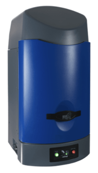
- High efficiency in vivo and ex vivo imaging
- Compatible with 5-mouse anesthesia platform
- High-quality optical luminescence and fluorescence imaging
- Equipped with Aura imaging software 4.0
- Smart exposure captures both faint and strong signals in a single image
- Features to prevent overexposure from individual animal readings
- Smooth setting allows maximum sensitivity for clear, non-pixelated images
- Variety of emission and excitation filters
- Advanced data software for comprehensive drug progression illustrations in longitudinal studies
Learn more about our Imaging Services for Preclinical Evaluation.
Contact our experts to help advance your orthotopic tumor models with our trusted preclinical research services.

Key Features of TD2’s Imaging Services:
Our imaging capabilities provide a robust platform for understanding tumor biology and treatment responses. Key features include:
- High-resolution imaging for precise evaluation of tumor growth and metastasis
- Longitudinal imaging for tracking treatment efficacy over time
- Detailed visualization of biological processes in live animal models
- Advanced data analysis for comprehensive assessment of therapeutic impact
TD2’s imaging services are designed to address the most critical questions in your preclinical development. By leveraging our imaging technology, you can enhance the accuracy and relevance of your preclinical studies, ultimately facilitating the transition to clinical trials. Our capabilities provide precise and actionable data, ensuring that your research is grounded in robust and reliable information.
Figure 1: Site and cell lines available for orthotopic implantation.

Comprehensive Preclinical Services
Regardless of the development question, TD2 has the specialized model you need to move your drug forward. Clients have access to humanized models, induction models, as well as specialty surgical and orthotopic models. We specialize in all areas of oncology and work diligently to understand your therapeutics’ mechanism of action. We match that with clinical development strategies for an efficient path forward.
- Non-GLP Safety/Tolerability
- In vitro Pharmacology Assays
- DMPK/ADME
- Orthotopic and Imaging tumor models
- CAR-T and Adoptive Cell Therapy models
- Syngeneic mouse models
- Diet-Induced Obesity Tumor Model
- Humanized Immune Checkpoint Inhibitor Mouse Models
- Metabolomics and Proteomics Analysis
- Flow Cytometry