Immunophenotyping
Immunophenotyping is a tool used to profile immune cells in peripheral blood, tissues and tumor samples. Combinations of antibodies, tagged with different fluorochomes, are used to detect proteins on the cell surface or within the cell (intracellular). Labeled cells are then acquired on a flow cytometer. TD2 has extensive experience designing immunophenotyping panels, from standard panels, to more complex panels that include activation markers, exhaustion markers, intracellular cytokines, and phosphorylation of intracellular proteins. We currently support immunophenotyping across multiple species, including human, non-human primate, canine, pig, mouse and rat.
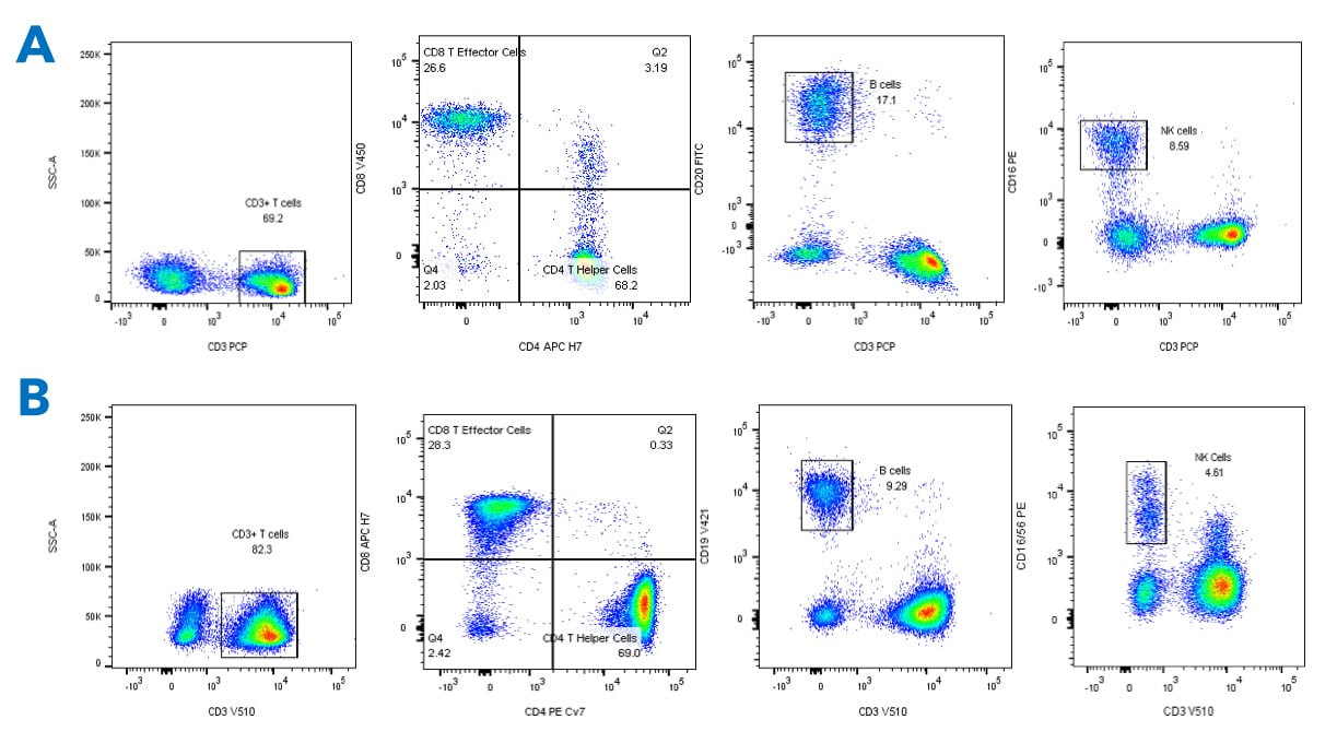
For example, standard TBNK panels are gated through CD45+ Lymphocytes to CD3+ cells, and then to CD4 (T Helper) and CD8 (T Effector) populations. B cells are identified as CD3-CD20+ and NK cells as CD3-CD16+. Examples of Non-Human Primate (A) and Human (B) TBNK Panels are shown below.
Request more information about Immunophenotyping
Contact our experts to help advance your drug development with TD2's trusted Flow Cytometry Services.
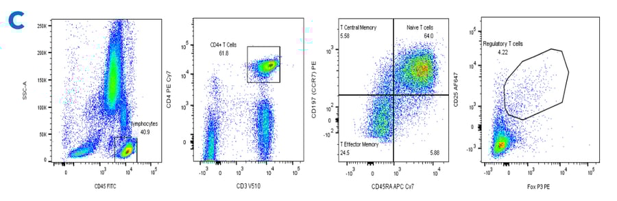
We have the ability to measure 10-colors/parameters within a single tube. We can further indentify T cell subsets (Central Memory, Effector Memory, Regulatory T cells, Exhausted T cells). For example, in panel C, Human CD4 cells are gated from CD45+ Lymphocytes, and further gated by CD45RA vs CCR7 to define memory subsets, or to Regulatory T cells.
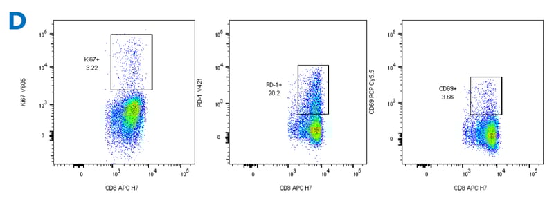
In addition, the functional/activation status of these T cell subsets can be measured: in Panel D, Ki67 marks proliferation in CD8 cells, and PD-1 and CD69 are used to assess activation. Only CD8 cells are shown, but the same analysis can be applied to CD4 cells.
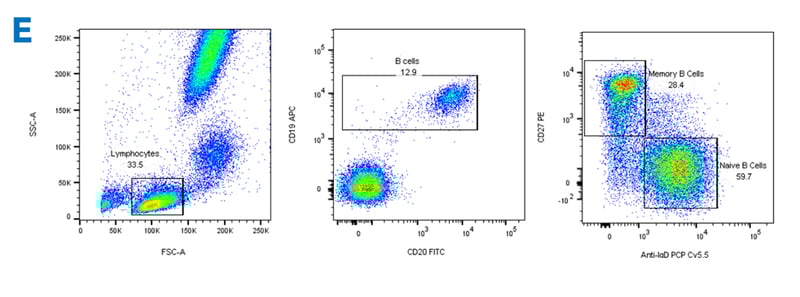
But, it’s not all about T cells: Immunophenotyping can be used to identify Subsets of B cells. In Panel E, from the lymphocyte gate, B cells are gated by CD19 vs CD20. CD19+ cells are further gated to Memory and Naïve B cell subsets. Plasma cells can also be quantitated With the Addition of CD38 to the panel.
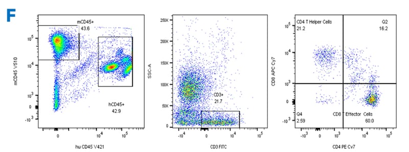
Humanized mice are genetically immune-deficient mice that have been engrafted with human stem cells to reconstitute a human immune system. They provide an invaluable tool for studying human immune cell function and the effects of drugs on human immune cells. Immunophenotyping can be done by first separating human leukocytes (hCD45+) from existing mouse leukocytes (mCD45+) Next, T cells are gated by CD3, and finally, CD4 and CD8 populations determined. Although not shown, further subsets of T cells and their activation/proliferation can be determined.
Immunophenotyping is an essential tool for monitoring drug treatment, or disease status and progression.
We are pleased to offer the following immunophenotyping panels.
- Standard T cell, B Cell and NK Panels (TBNK)
- Regulatory T cells
- Memory, Naïve and Effector T cells
- TH1/TH2/TH17/TH9 T cells
- T cell activation/proliferation
- Intracellular Cytokines
- Phospho pERK, pAKT, pSTAT1, pSTAT5
- Memory B cells and Plasma Cells
- Humanized Mouse Immunophenotyping
- Customized Immunophenotyping Panels
TD2 is also excited to announce the addition of Cytek Aurora instrumentation to expand our antibody panel configurations to 20+ colors. These expanded panels are available for use on exploratory projects.
Additional Resources


Regulatory, Clinical, Preclinical, PK/ADME, Flow
Comprehensive Oncology Ecosystem


![]() Receptor Occupancy
Receptor Occupancy
Binding of fluorophore labeled “drug” in relation to decreasing dose of unlabeled “drug”

![]() Receptor Occupancy
Receptor Occupancy
![]() PBMC Services
PBMC Services
Evaluate responses of PBMCs by profiling the function/phenotype of subpopulations

![]() Immune Population Phenotyping
Immune Population Phenotyping
Immune cell population analysis of subcutaneous CT26 murine colon carcinoma tumors
CT26 SC tumors: Gating strategy T cells and NK cells.
Gating involved identifying the total cell population, and then gate out the doublets, gate out the dead cells, move on to our first marker CD45 which gates out any non-immune cells. The bottom right cytogram shows the different NK cell populations. The far left middle cytogram depicts the two T cell population CD4+ and CD8+. Values are % of total CD45+ cells.
![]() Cancer Cells/Tumor Specific Marker Expression
Cancer Cells/Tumor Specific Marker Expression
Cancer cells/tumors specific markers expression
- Viability dye inclusion
- Cell lines and single-cell suspension tumors
- Surface staining

![]() Fluorescent Proteins Expression
Fluorescent Proteins Expression
Fluorescent proteins expression
- Viability dye inclusion
- Cell lines and transfected primary cells
- Surface staining

![]() Cell Cycle Analysis
Cell Cycle Analysis
Cell cycle analysis A549 cells
- Propidium iodide
- Cell lines

![]() Intracellular Protein Analysis
Intracellular Protein Analysis
- Intracellular Cytokines
- Phosphorylated Proteins
- T-Regs
- Phosphoroteins
- Cytokines

![]() Cellular Functional Assays
Cellular Functional Assays
- NK Cell Function
- Basophil Activation Test (BAT)
