---
title: Advanced Preclinical Flow Cytometry Analysis Services
description: Discover TD2's advanced preclinical flow cytometry services for oncology research. Our platform offers comprehensive immunophenotyping, drug mechanism analysis, and high-throughput assays designed to optimize your drug's therapeutic impact. Tailor your treatment strategies with our expert guidance and cutting-edge technology.
image: https://td2inc.com/hubfs/Assets/Images/og-home.jpg
---
×
[](https://td2inc.com/?hsLang=en)

[](https://td2inc.com/?hsLang=en)

# Advanced Preclinical Flow Cytometry Analysis Services
### Precision Immunophenotyping & Drug Impact Analysis for Oncology Research
Gain a deeper understanding of your drug’s therapeutic effects through high-throughput flow cytometry analysis across a diverse array of assays and cell types. TD2’s cutting-edge platform offers comprehensive immunophenotyping, enabling you to delve into your drug’s mechanism of action and pharmacodynamics, ultimately strengthening the efficacy of your immunotherapy while effectively minimizing unwanted side effects.
[View Our Clinical Flow Cytometry Solutions](https://td2inc.com/clinical-flow-cytometry?hsLang=en)
**Features & Benefits:**
- **In-Depth Analysis:** Explore immune composition within vital organs such as tumors, spleen, and lymph nodes, allowing you to tailor treatment strategies for maximum impact.
- **Precise Impact Assessment:** Evaluate the therapy’s influence on the frequencies and functions of specific cell subsets, optimizing its performance.
- **Expert Guidance:** Access our dedicated FACS team’s expertise to customize protocols, panel configurations, gating strategies, and more, ensuring the most relevant results for your study.
- **Efficient High-Throughput:** Characterize cells with ease, using minimal sample volumes, thanks to our advanced equipment, including Miltenyi gentleMACS™ Octo Dissociator, MultiMACS Cell24 Separator, Miltenyi MACSQuant Analyzer 16, BD FACSCanto™, BD FACSCanto™ II, 10-color BD FACSCanto, Cytek Aurora, and Millipore Guava PCA
With TD2’s flow cytometry platform, you can confidently navigate the complexities of immune phenotyping and rare cell populations, gaining valuable insights into your drug’s potential and optimizing its therapeutic impact
[Flow Cytometry FAQ](https://td2inc.com/flow-cytometry-faq?hsLang=en)
### Request more information about our Flow Cytometry Services
Contact our experts to help advance your drug development with TD2's trusted Flow Cytometry Services.
### Capabilities and Assays Include:
**Tissue Dissociation/Single-Cell Suspension**
- Spleen, Tumors, Lymph nodes
[**Receptor Occupancy**](https://td2inc.com/receptor-occupancy?hsLang=en)x
[**PBMC Services**](https://td2inc.com/peripheral-blood-mononuclear-cells?hsLang=en)x
**Primary Cell Isolation**
- Pan T cells isolation kit, mouse
- CD4+ T cell isolation kit, mouse
- isolation of untouched mouse cells from single-cell suspensions from spleen
[**Immune Population Phenotyping**](https://td2inc.com/immunophenotyping?hsLang=en)x
- Panel design 20+ colors
- Whole blood, bone marrow, spleen, and tumor
- Viability dye inclusion
- Surface staining
- Intracellular staining, including:
- Cytoplasmic proteins
- Phospo-proteins (specific buffer set)
- Nuclear transcription factors (specific buffer set)
- Cytokines (specific buffer set)
- T-lymphocyte, B-lymphocyte and Natural Killer cells (TBNK)
- T Regulatory Cells
- T Memory, Naive and Effector Cells
- T Cell Activation
- B Memory and Plasma cells
- Humanized mice panels
- Custom panels
- CAP TBNK Panel
**Cancer Cells/Tumor Specific Marker Expression**x
- Viability dye inclusion
- Cell lines and single-cell suspension tumors
- Surface staining
**Fluorescent Proteins Expression**x
- Viability dye inclusion
- Cell lines and transfected primary cells
- Surface staining
**Cell Cycle Analysis**x
- Propidium iodide
- Cell lines
- Solid tumors (under development)
**Intracellular Protein Analysis**x
- Intracellular Cytokines
- Phosphorylated Proteins
[**Cellular Functional Assays**](https://td2inc.com/functional-assays?hsLang=en)x
- NK Cell Function
- Basophil Activation Test (BAT)
## Additional Resources
[](https://td2inc.com/wp-optimizing-specimen-stability-a-comparative-analysis-of-shipping-protocols?hsLang=en)
Flow
## [Optimizing Specimen Stability – A Comparative Analysis of Shipping Protocols](https://td2inc.com/wp-optimizing-specimen-stability-a-comparative-analysis-of-shipping-protocols?hsLang=en)
[](https://blog.td2inc.com/leveraging-flow-cytometry-to-drive-better-patient-outcomes-in-hematologic-malignancy-clinical-trials?hsLang=en)
Clinical
## [Leveraging Flow Cytometry to Drive Better Patient Outcomes in Hematologic Malignancy Clinical Trials](https://blog.td2inc.com/leveraging-flow-cytometry-to-drive-better-patient-outcomes-in-hematologic-malignancy-clinical-trials?hsLang=en)
[](https://td2inc.com/lp-fcsl-poster-2024-immunophenotyping-pig-immune-cells-by-flow-cytometry-a-powerful-tool-for-pharmacology-and-toxicology-studies/?hsLang=en)
Flow
## [Immunophenotyping Pig Immune Cells by Flow Cytometry: A Powerful Tool for Pharmacology and Toxicology Studies](https://td2inc.com/lp-fcsl-poster-2024-immunophenotyping-pig-immune-cells-by-flow-cytometry-a-powerful-tool-for-pharmacology-and-toxicology-studies/?hsLang=en)
[](https://td2inc.com/lp-fcsl-poster-2024-flow-cytometry-application-in-water-microbiology/?hsLang=en)
Flow
## [Flow Cytometry Application in Water Microbiology: Testing Bacterial Content in Water](https://td2inc.com/lp-fcsl-poster-2024-flow-cytometry-application-in-water-microbiology/?hsLang=en)
[](https://td2inc.com/lp-fcsl-poster-2024-flow-cytometry-processing-and-analysis-of-tumor-specimens/?hsLang=en)
Flow
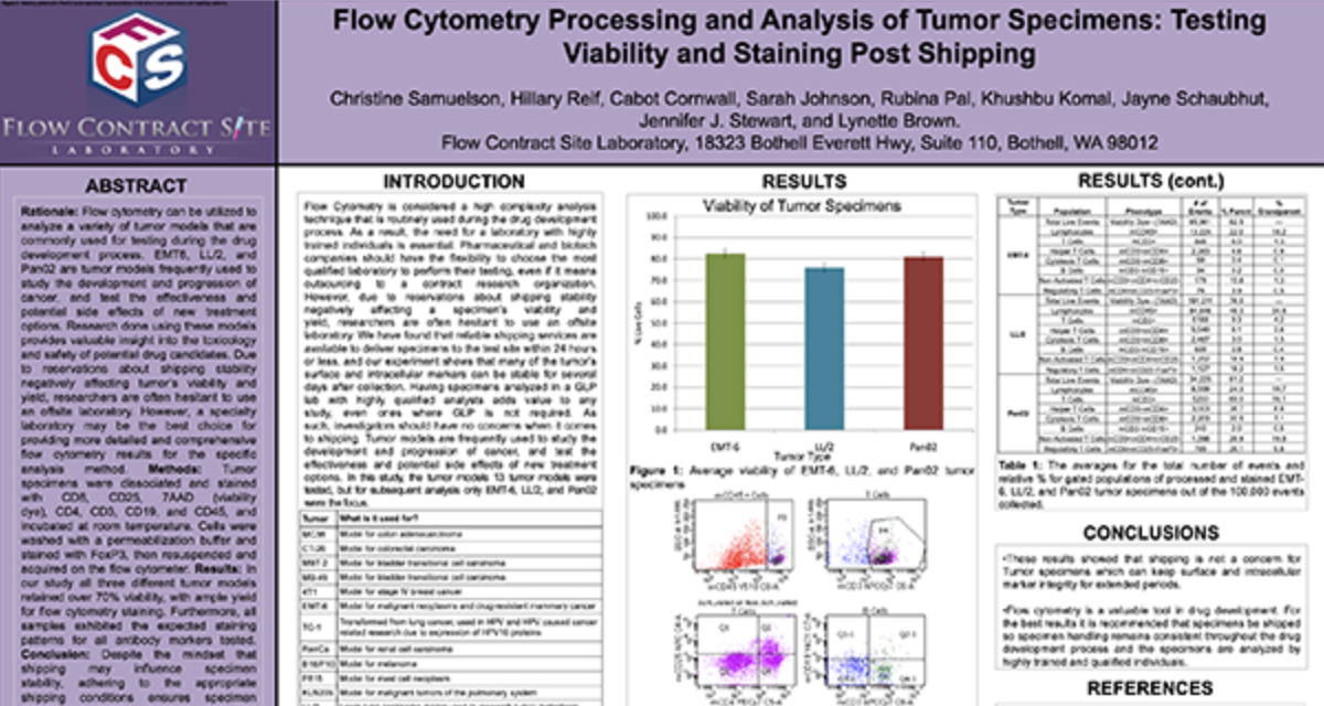
## [Flow Cytometry Processing and Analysis of Tumor Specimens: Testing Viability and Staining Post Shipping](https://td2inc.com/lp-fcsl-poster-2024-flow-cytometry-processing-and-analysis-of-tumor-specimens/?hsLang=en)
[](https://td2inc.com/lp-fcsl-poster-2024-flow-cytometry-based-assessment-of-innate-immune-function-using-phagocytosis-and-reactive-oxygen-species-measurement/?hsLang=en)
Flow
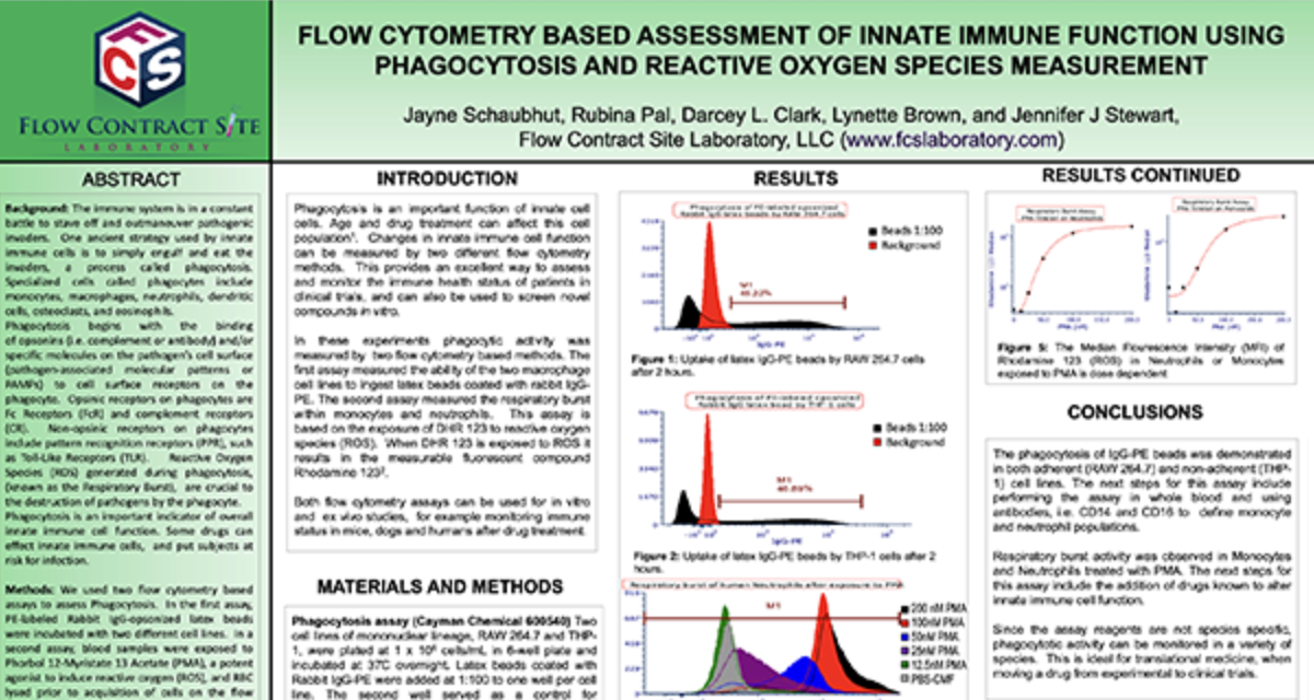
## [Flow Cytometry Based Assessment of Innate Immune Function Using Phagocytosis and Reactive Oxygen Species Measurement](https://td2inc.com/lp-fcsl-poster-2024-flow-cytometry-based-assessment-of-innate-immune-function-using-phagocytosis-and-reactive-oxygen-species-measurement/?hsLang=en)
[](https://td2inc.com/lp-fcsl-poster-2024-measurement-of-cytokine-mrna-expression/?hsLang=en)
Flow
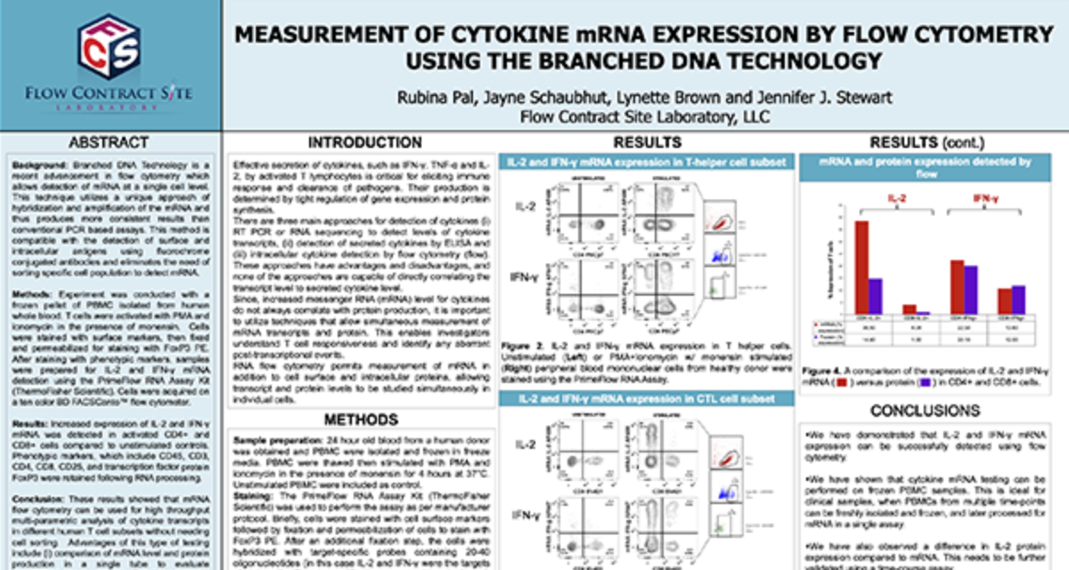
## [Measurement of Cytokine mRNA Expression by Flow Cytometry Using the Branched DNA Technology](https://td2inc.com/lp-fcsl-poster-2024-measurement-of-cytokine-mrna-expression/?hsLang=en)
[](https://td2inc.com/lp-fcsl-poster-2024-10-color-flow-cytometry-panels-for-the-evaluation-of-t-cell-subsets/?hsLang=en)
Flow
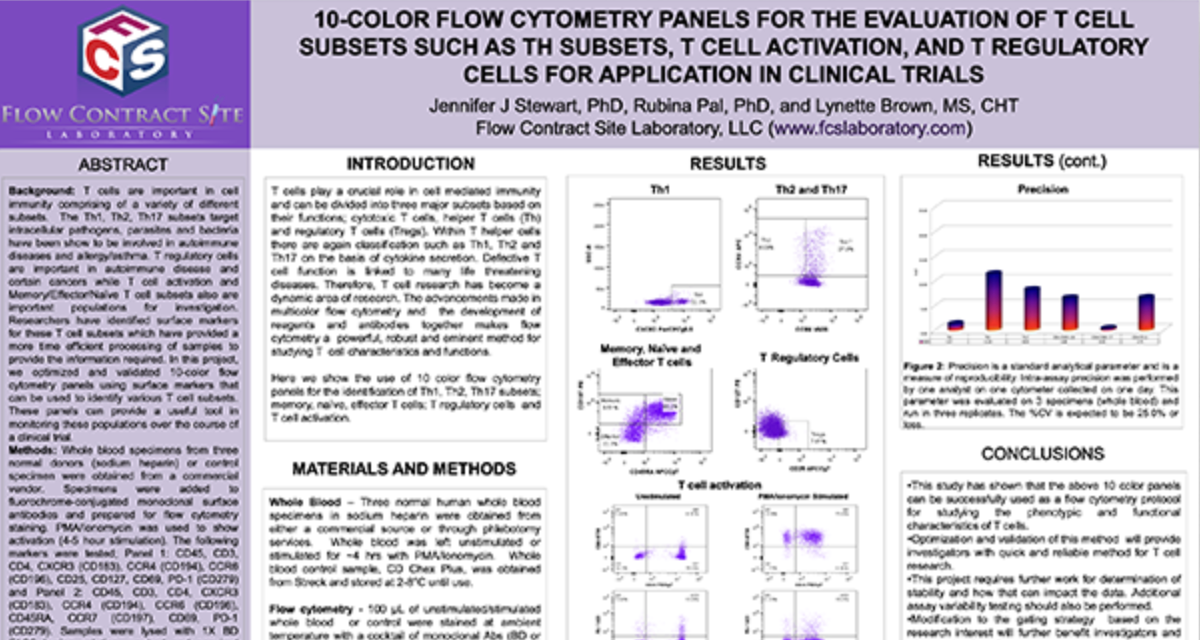
## [10-Color Flow Cytometry Panels for the Evaluation of T Cell Subsets Such as TH Subsets, T Cell Activation, and T Regulatory Cells for Application in Clinical Trials](https://td2inc.com/lp-fcsl-poster-2024-10-color-flow-cytometry-panels-for-the-evaluation-of-t-cell-subsets/?hsLang=en)
[](https://td2inc.com/lp-fcsl-poster-2024-the-advantages-of-fluorescent-barcoding-of-human-cells-using-flow-cytometry/?hsLang=en)
Flow
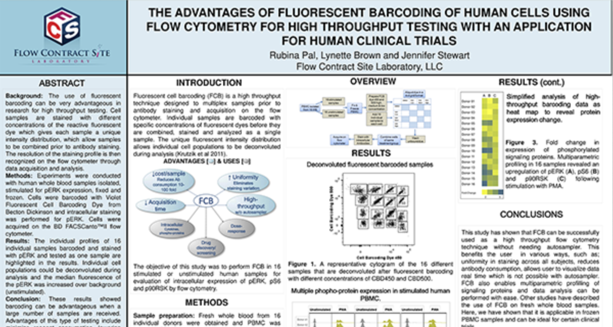
## [The Advantages of Fluorescent Barcoding of Human Cells Using Flow Cytometry for High Throughput Testing With an Application For Human Clinical Trials](https://td2inc.com/lp-fcsl-poster-2024-the-advantages-of-fluorescent-barcoding-of-human-cells-using-flow-cytometry/?hsLang=en)
[](https://td2inc.com/lp-fcsl-poster-2024-the-activation-profiling-of-stat-in-human-whole-blood-samples/?hsLang=en)
Flow
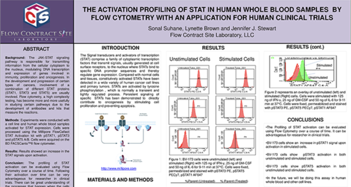
## [The Activation Profiling of Stat in Human Whole Blood Samples By Flow Cytometry With an Application for Human Clinical Trials](https://td2inc.com/lp-fcsl-poster-2024-the-activation-profiling-of-stat-in-human-whole-blood-samples/?hsLang=en)
Load More
## GET STARTED
## Work with a team who believes in your research as much as you do.
Are you ready to start your Flow Cytometry studies? Partner with a collaborative oncology CRO that believes in your treatment as much as you do. Take the first step today and contact our experts.
[Get Started Now](https://td2inc.com/contact-us/?hsLang=en)
×
**Receptor Occupancy**
### Binding of fluorophore labeled “drug” in relation to decreasing dose of unlabeled “drug”
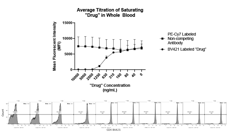
Receptor Occupancy
×
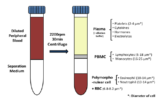
**PBMC Services**
### Evaluate responses of PBMCs by profiling the function/phenotype of subpopulations
×
**Immune Population Phenotyping**
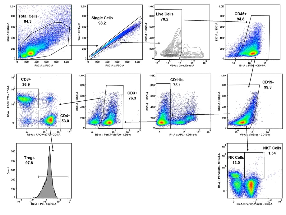
### Immune cell population analysis of subcutaneous CT26 murine colon carcinoma tumors
CT26 SC tumors: Gating strategy T cells and NK cells.
Gating involved identifying the total cell population, and then gate out the doublets, gate out the dead cells, move on to our first marker CD45 which gates out any non-immune cells. The bottom right cytogram shows the different NK cell populations. The far left middle cytogram depicts the two T cell population CD4+ and CD8+. Values are % of total CD45+ cells.
×
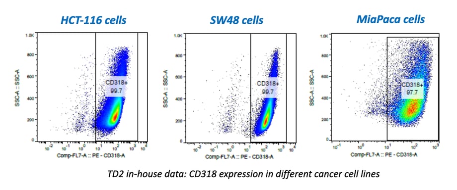
**Cancer Cells/Tumor Specific Marker Expression**
### Cancer cells/tumors specific markers expression
- Viability dye inclusion
- Cell lines and single-cell suspension tumors
- Surface staining
×
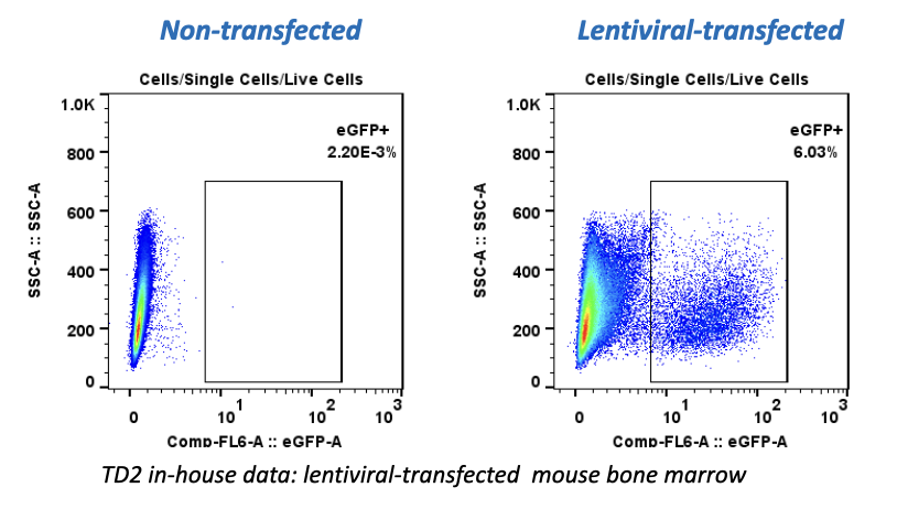
**Fluorescent Proteins Expression**
### Fluorescent proteins expression
- Viability dye inclusion
- Cell lines and transfected primary cells
- Surface staining
×
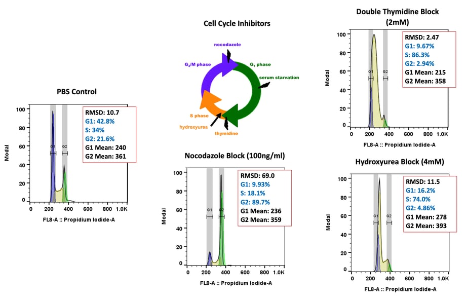
**Cell Cycle Analysis**
### Cell cycle analysis A549 cells
- Propidium iodide
- Cell lines
×
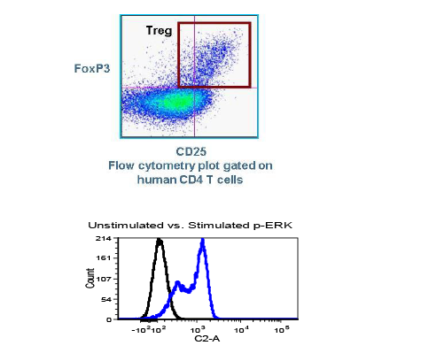
**Intracellular Protein Analysis**
- Intracellular Cytokines
- Phosphorylated Proteins
- T-Regs
- Phosphoroteins
- Cytokines
×
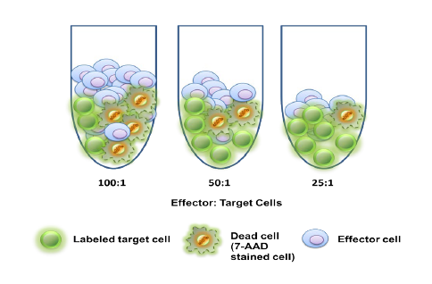
**Cellular Functional Assays**
- NK Cell Function
- Basophil Activation Test (BAT)
[](https://td2inc.com/?hsLang=en)
Office
13208 E. Shea Blvd,
Suite 100
Scottsdale, AZ 85259
Phone
(602) 358-8300
Connect with us
-
-

© 2026 Translational Drug Development. All Rights Reserved. [Terms and Conditions](https://td2inc.com/terms-and-conditions?hsLang=en) | [Privacy Policy](https://td2inc.com/privacy-policy?hsLang=en)