---
title: Peripheral Blood Mononuclear Cells (PBMC)
description: At TD2 we have extensive experience in PBMC isolation and cryopreservation. We can help your next PBMC projects with ensuring high cell viability and recovery, timely, standardized and quality results.
image: https://td2inc.com/hubfs/Assets/Images/og-home.jpg
---
×
[](https://td2inc.com/?hsLang=en)

[](https://td2inc.com/?hsLang=en)

# Peripheral Blood Mononuclear Cells (PBMC)
The separation of Peripheral Blood Mononuclear Cells (PBMC) may be required for certain applications including flow cytometry. At FCSL we can provide support for PBMC separation as required for your specific assay. Please inquire to start the scientific discussion.
Most flow cytometry assays can be performed on whole blood or on samples that have been drawn and undergone further processing. For some applications it may be ideal to prepare processed whole blood in order to store cells for longer periods than fresh blood or to perform batch testing for each subject over several time points. In these instances, peripheral blood mononuclear cells (PBMCs) -are prepared and frozen. PBMCs consist of lymphocytes (T cells, B cells& NK cells), monocytes and dendritic cells. In humans, lymphocytes make up the majority of the PBMC population, followed by monocytes and only a small percentage of dendritic cells PBMCs are the critical component of the immune system that fights infections and diseases. Therefore, key immune responses can be evaluated using PBMCs by profiling - the function and/or phenotype of the different PBMC subpopulations.
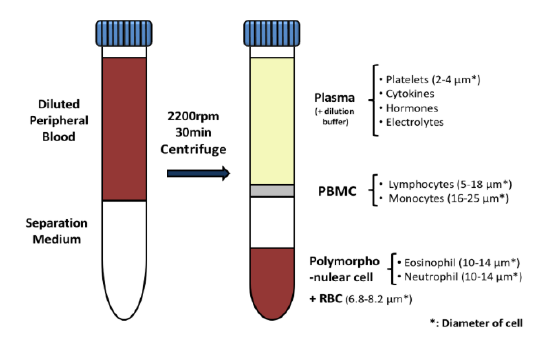
PBMCs must be separated from whole blood. At TD2, we -use density gradient separation for most of our PBMC preparations.

**Figure 1.** PBMC isolation by Density gradient centrifugation
### Request more information about Peripheral Blood Mononuclear Cells
Contact our experts to help advance your drug development with TD2's trusted Flow Cytometry Services.
As shown in Figure 1, each cell population exhibits a unique migration pattern through the medium and each layer contains specific blood components. The cells are isolated by extracting the respective layer. Isolated PBMCs are washed and then evaluated for cell viability and cell count - using the Guava PCA (Personal Cell Analysis) as shown in Figure 2 and 3.

**Figure 2.** Guava PCA flow Cytometer

**Figure 3.** Cell viability and viable cell count by Guava PCA
Viable PBMCs are cryopreserved in freezing media. Our laboratory has both liquid nitrogen and -80° freezers for short term storage until the specific flow cytometry assay can be performed or shipped to other laboratories for other downstream applications.
At TD2 we have extensive experience in PBMC isolation and cryopreservation. We can help your next PBMC projects with ensuring high cell viability and recovery, timely, standardized and quality results. Please inquire to start the scientific discussion.
## Additional Resources
[](https://td2inc.com/poster-development-of-a-receptor-occupancy-assay-for-a-therapeutic-bi-specific-antibody?hsLang=en)
Flow
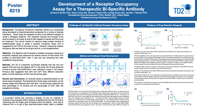
## [Development of a Receptor Occupancy Assay for a Therapeutic Bi-Specific Antibody](https://td2inc.com/poster-development-of-a-receptor-occupancy-assay-for-a-therapeutic-bi-specific-antibody?hsLang=en)
[](https://td2inc.com/poster-measurement-of-basophil-activation-using-a-flow-cytometry-method-for-applications-in-clinical-research-and-clinical-trials?hsLang=en)
Flow
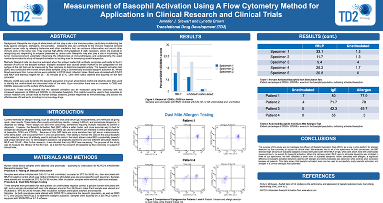
## [Measurement of Basophil Activation Using a Flow Cytometry Method for Applications in Clinical Research and Clinical Trials](https://td2inc.com/poster-measurement-of-basophil-activation-using-a-flow-cytometry-method-for-applications-in-clinical-research-and-clinical-trials?hsLang=en)
[](https://td2inc.com/wp-optimizing-specimen-stability-a-comparative-analysis-of-shipping-protocols?hsLang=en)
Flow
## [Optimizing Specimen Stability – A Comparative Analysis of Shipping Protocols](https://td2inc.com/wp-optimizing-specimen-stability-a-comparative-analysis-of-shipping-protocols?hsLang=en)
[](https://td2inc.com/sitc-2024-poster-design-and-validation-of-a-model-of-a-receptor-occupancy-assay?hsLang=en)
Flow
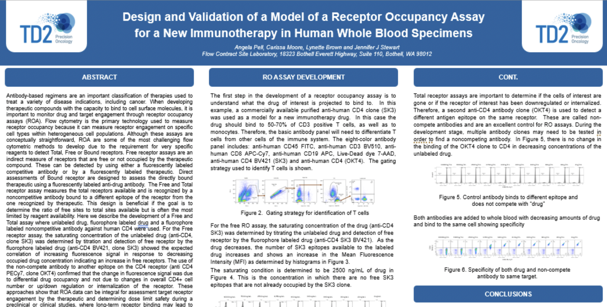
## [Design and Validation of a Model of a Receptor Occupancy Assay for a New Immunotherapy in Human Whole Blood Specimens](https://td2inc.com/sitc-2024-poster-design-and-validation-of-a-model-of-a-receptor-occupancy-assay?hsLang=en)
[](https://td2inc.com/welcome-to-td2s-comprehensive-oncology-ecosystem?hsLang=en)
Regulatory, Clinical, Preclinical, PK/ADME, Flow
## [Comprehensive Oncology Ecosystem](https://td2inc.com/welcome-to-td2s-comprehensive-oncology-ecosystem?hsLang=en)
[](https://td2inc.com/wp-flow-cytometry-based-receptor-occupancy-assays?hsLang=en)
Flow
## [Flow Cytometry-Based Receptor Occupancy Assays: Technical Challenges and Applications in Drug Development](https://td2inc.com/wp-flow-cytometry-based-receptor-occupancy-assays?hsLang=en)
[](https://td2inc.com/aacr-2024-poster-510?hsLang=en)
Flow
## [Flow cytometry evaluation of cell cycle arrest in A549 human non-small cell lung cancer cells and tumors following treatment with cell cycle inhibitors: A multifaceted approach to targeting cancer](https://td2inc.com/aacr-2024-poster-510?hsLang=en)
[](https://td2inc.com/lp-fcsl-poster-2024-immunophenotyping-pig-immune-cells-by-flow-cytometry-a-powerful-tool-for-pharmacology-and-toxicology-studies/?hsLang=en)
Flow
## [Immunophenotyping Pig Immune Cells by Flow Cytometry: A Powerful Tool for Pharmacology and Toxicology Studies](https://td2inc.com/lp-fcsl-poster-2024-immunophenotyping-pig-immune-cells-by-flow-cytometry-a-powerful-tool-for-pharmacology-and-toxicology-studies/?hsLang=en)
[](https://td2inc.com/lp-fcsl-poster-2024-flow-cytometry-application-in-water-microbiology/?hsLang=en)
Flow
## [Flow Cytometry Application in Water Microbiology: Testing Bacterial Content in Water](https://td2inc.com/lp-fcsl-poster-2024-flow-cytometry-application-in-water-microbiology/?hsLang=en)
[](https://td2inc.com/lp-fcsl-poster-2024-flow-cytometry-processing-and-analysis-of-tumor-specimens/?hsLang=en)
Flow
## [Flow Cytometry Processing and Analysis of Tumor Specimens: Testing Viability and Staining Post Shipping](https://td2inc.com/lp-fcsl-poster-2024-flow-cytometry-processing-and-analysis-of-tumor-specimens/?hsLang=en)
[](https://td2inc.com/lp-fcsl-poster-2024-flow-cytometry-based-assessment-of-innate-immune-function-using-phagocytosis-and-reactive-oxygen-species-measurement/?hsLang=en)
Flow
## [Flow Cytometry Based Assessment of Innate Immune Function Using Phagocytosis and Reactive Oxygen Species Measurement](https://td2inc.com/lp-fcsl-poster-2024-flow-cytometry-based-assessment-of-innate-immune-function-using-phagocytosis-and-reactive-oxygen-species-measurement/?hsLang=en)
[](https://td2inc.com/lp-fcsl-poster-2024-measurement-of-cytokine-mrna-expression/?hsLang=en)
Flow
## [Measurement of Cytokine mRNA Expression by Flow Cytometry Using the Branched DNA Technology](https://td2inc.com/lp-fcsl-poster-2024-measurement-of-cytokine-mrna-expression/?hsLang=en)
[](https://td2inc.com/lp-fcsl-poster-2024-10-color-flow-cytometry-panels-for-the-evaluation-of-t-cell-subsets/?hsLang=en)
Flow
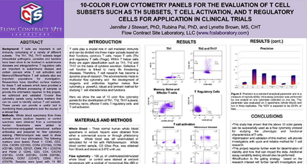
## [10-Color Flow Cytometry Panels for the Evaluation of T Cell Subsets Such as TH Subsets, T Cell Activation, and T Regulatory Cells for Application in Clinical Trials](https://td2inc.com/lp-fcsl-poster-2024-10-color-flow-cytometry-panels-for-the-evaluation-of-t-cell-subsets/?hsLang=en)
[](https://td2inc.com/lp-fcsl-poster-2024-the-advantages-of-fluorescent-barcoding-of-human-cells-using-flow-cytometry/?hsLang=en)
Flow
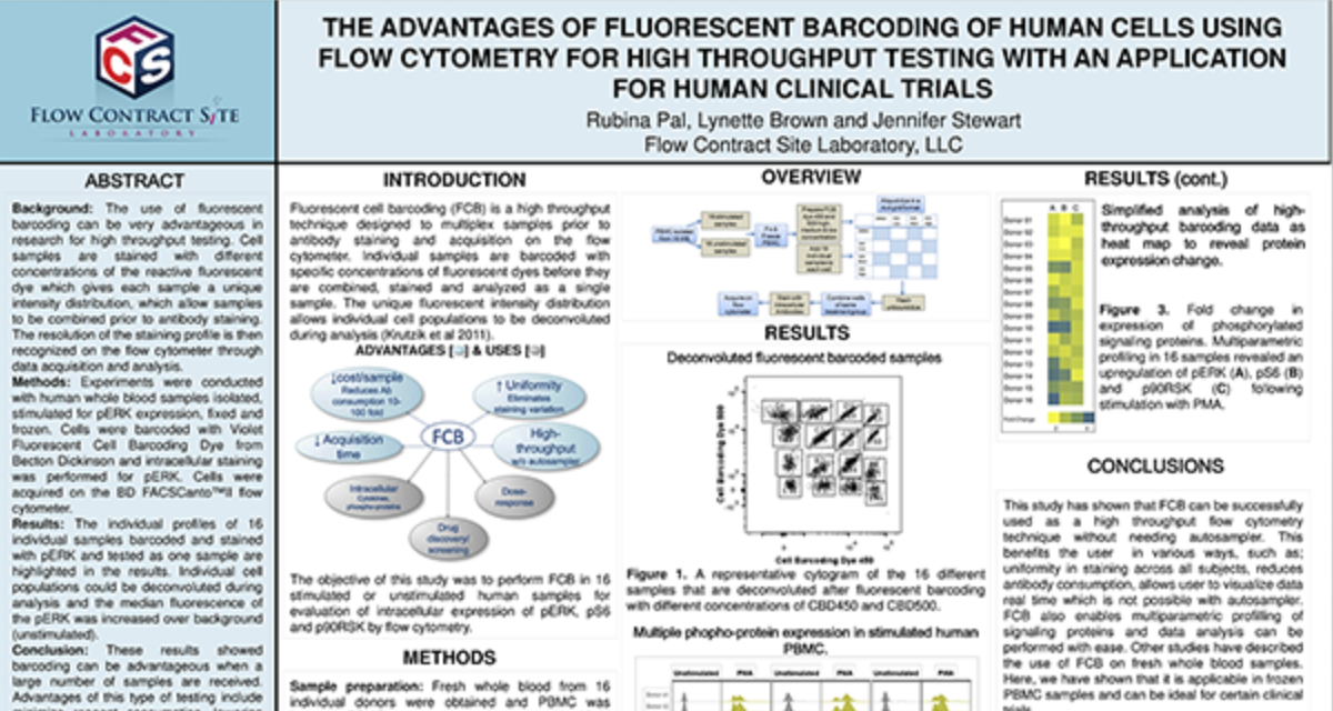
## [The Advantages of Fluorescent Barcoding of Human Cells Using Flow Cytometry for High Throughput Testing With an Application For Human Clinical Trials](https://td2inc.com/lp-fcsl-poster-2024-the-advantages-of-fluorescent-barcoding-of-human-cells-using-flow-cytometry/?hsLang=en)
[](https://td2inc.com/lp-fcsl-poster-2024-the-activation-profiling-of-stat-in-human-whole-blood-samples/?hsLang=en)
Flow
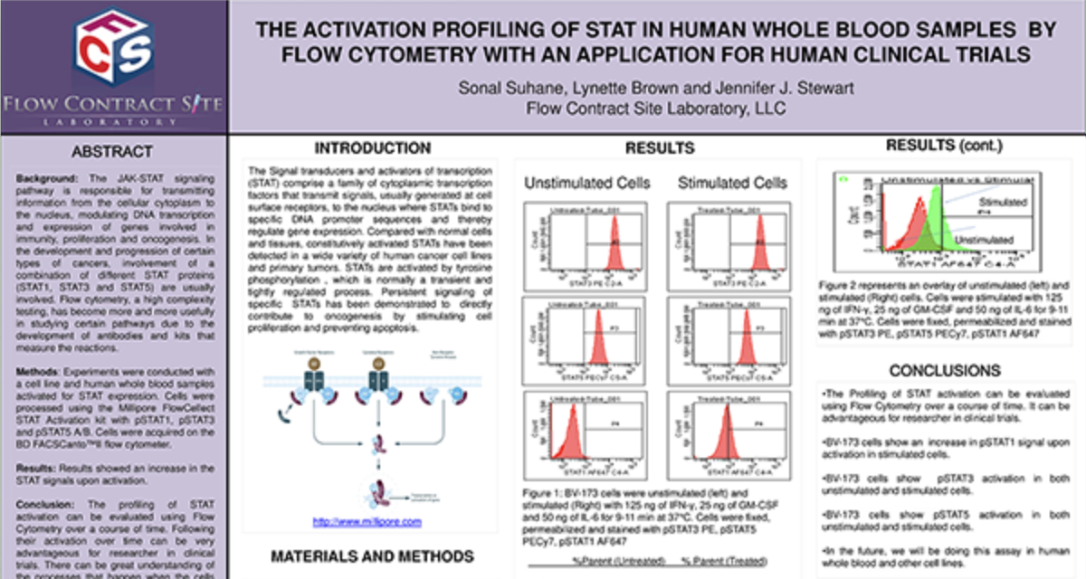
## [The Activation Profiling of Stat in Human Whole Blood Samples By Flow Cytometry With an Application for Human Clinical Trials](https://td2inc.com/lp-fcsl-poster-2024-the-activation-profiling-of-stat-in-human-whole-blood-samples/?hsLang=en)
[](https://td2inc.com/lp-fcsl-poster-2024-design-and-validation-of-a-model-of-a-receptor-occupancy-assay/?hsLang=en)
Flow
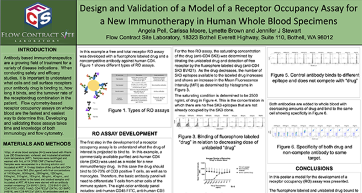
## [Design and Validation of a Model of a Receptor Occupancy Assay for a New Immunotherapy in Human Whole Blood Specimens](https://td2inc.com/lp-fcsl-poster-2024-design-and-validation-of-a-model-of-a-receptor-occupancy-assay/?hsLang=en)
Load More
## GET STARTED
## Work with a team who believes in your research as much as you do.
Are you ready to start your Flow Cytometry studies? Partner with a collaborative oncology CRO that believes in your treatment as much as you do. Take the first step today and contact our experts.
[Get Started Now](https://td2inc.com/contact-us/?hsLang=en)
×
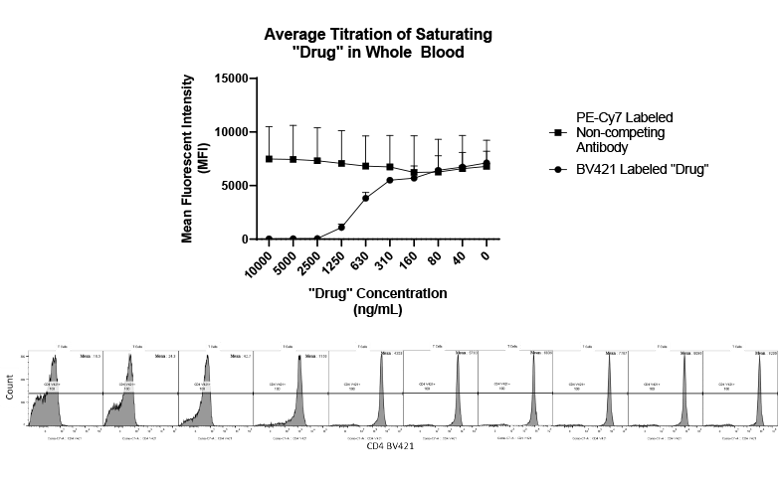
**Receptor Occupancy**
### Binding of fluorophore labeled “drug” in relation to decreasing dose of unlabeled “drug”
Receptor Occupancy
×
**PBMC Services**
### Evaluate responses of PBMCs by profiling the function/phenotype of subpopulations
×
**Immune Population Phenotyping**
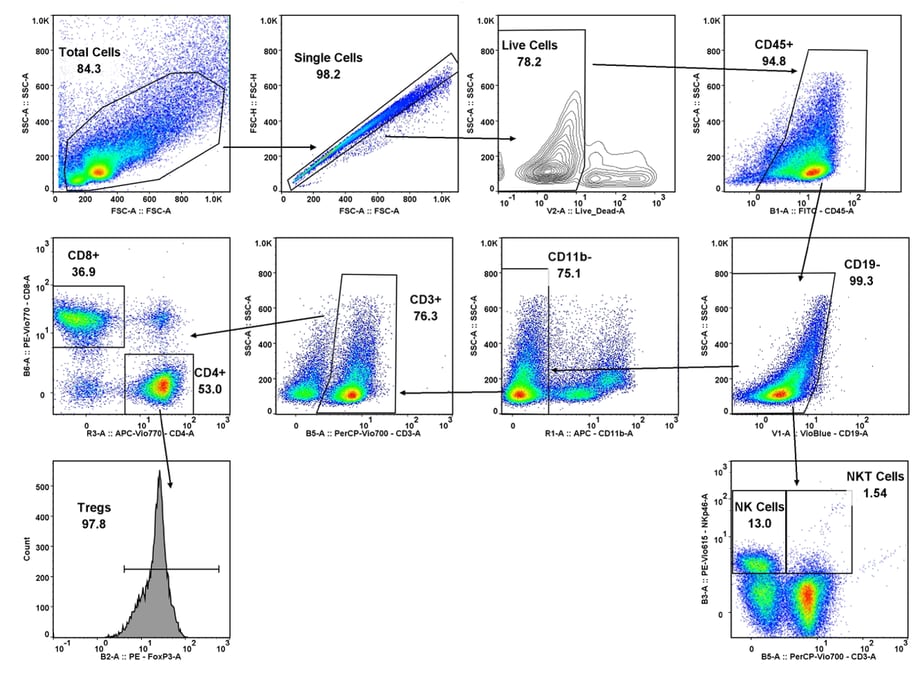
### Immune cell population analysis of subcutaneous CT26 murine colon carcinoma tumors
CT26 SC tumors: Gating strategy T cells and NK cells.
Gating involved identifying the total cell population, and then gate out the doublets, gate out the dead cells, move on to our first marker CD45 which gates out any non-immune cells. The bottom right cytogram shows the different NK cell populations. The far left middle cytogram depicts the two T cell population CD4+ and CD8+. Values are % of total CD45+ cells.
×
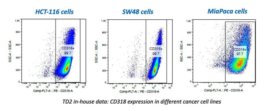
**Cancer Cells/Tumor Specific Marker Expression**
### Cancer cells/tumors specific markers expression
- Viability dye inclusion
- Cell lines and single-cell suspension tumors
- Surface staining
×
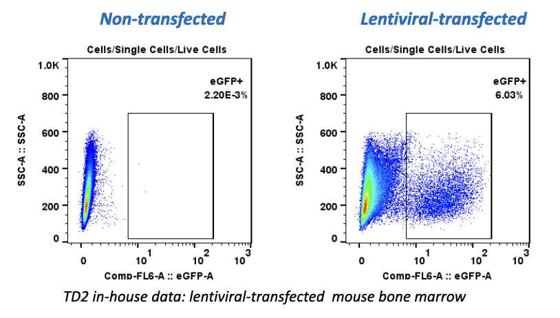
**Fluorescent Proteins Expression**
### Fluorescent proteins expression
- Viability dye inclusion
- Cell lines and transfected primary cells
- Surface staining
×
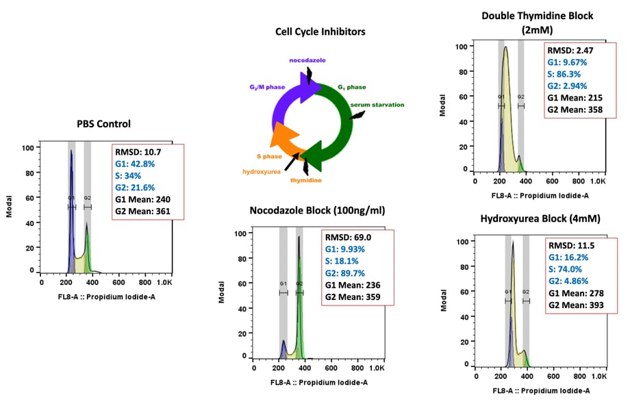
**Cell Cycle Analysis**
### Cell cycle analysis A549 cells
- Propidium iodide
- Cell lines
×
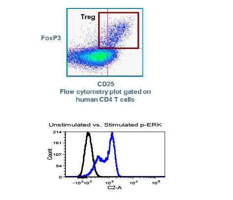
**Intracellular Protein Analysis**
- Intracellular Cytokines
- Phosphorylated Proteins
- T-Regs
- Phosphoroteins
- Cytokines
×
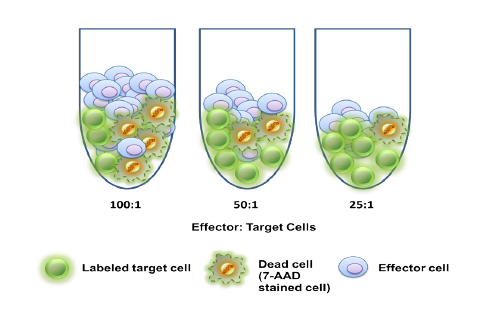
**Cellular Functional Assays**
- NK Cell Function
- Basophil Activation Test (BAT)
[](https://td2inc.com/?hsLang=en)
Office
13208 E. Shea Blvd,
Suite 100
Scottsdale, AZ 85259
Phone
(602) 358-8300
Connect with us
-
-

© 2026 Translational Drug Development. All Rights Reserved. [Terms and Conditions](https://td2inc.com/terms-and-conditions?hsLang=en) | [Privacy Policy](https://td2inc.com/privacy-policy?hsLang=en)